
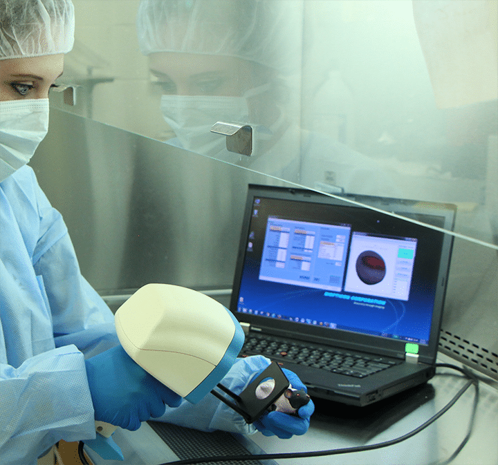
TumorImager 2TM is our next generation tumor scanner for subcutaneous tumor measurements on small lab animals. It uses a unique structured light tumor imaging system to capture both a 3D surface profile and a color image. The result is a one-of-a-kind tumor imaging providing a record of tumor shape and color. The patented algorithms can also isolate a tumor in the recorded image and calculate the volume or area. When interfaced to TumorManager 2TM this leads to a simple but robust, data-verified measurement system that offers speed, accuracy and flexibility.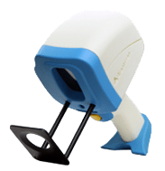
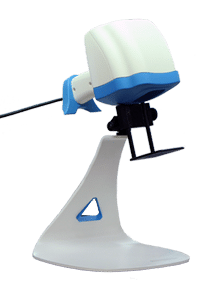
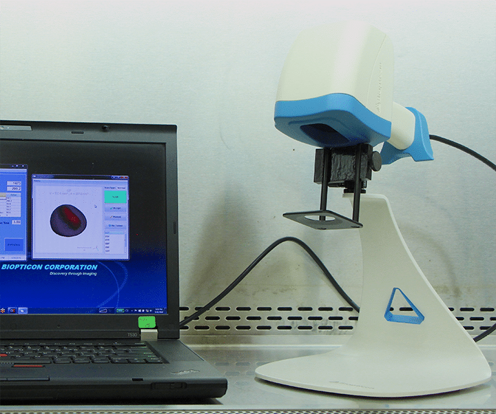
TumorImager 2TM can be quickly converted between a “supermarket-style” hand-held tumor scanner and hands-free scanner using the convenient multi-axis stand. No tools required! The whole unit can be placed in a biosafety hood and easily moved about the lab.
TumorImager 2TM constructs a 3D surface profile by projecting 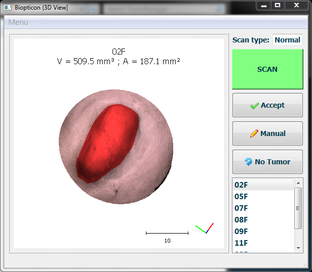 special light patterns on the animal. The relationship between different pattern images allows the creation of an animal surface profile. Like TumorImagerTM, our patented algorithms can automatically isolate a tumor within this profile. Once the tumor is located, volume, area, and other custom statistics are calculated. In addition, a color image and surface profile can be logged for future reference or analysis.
A typical animal scan takes just a few seconds:
TumorImager 2TM also easily allows for user-defined regions to be measured and to quickly initiate a rescan. This process generally takes less time than making caliper measurements for advanced tumor imaging.
| Scan modes | Hand-held or hands-free stand |
| Maximum tumor size | 30 mm |
| Maximum tumor height | 20 mm |
| Tumor scan time | <1 sec |
| Auto-Segmentation | ~3 sec |
| Manual Segmentation | Touchscreen, mouse |
| Resolution, z axis | 50µm |
| Computer Interface | USB 3.0 |
| Color Imaging format | 480×640 Color JPG |
| Surface profile format | OBJ, PLY |
| User Interfaces | Touch screen, mouse, foot pedal, and scanner button |